Skin Cancer Screening and ABCDE Melanoma Warning Signs
Key Points
- Common skin-cancer types include basal cell carcinoma, squamous cell carcinoma, and melanoma.
- Melanoma has the highest metastatic risk and requires early detection.
- Melanoma can occur in less sun-exposed areas (for example between toes or genital regions), so full-body surveillance teaching matters.
- Routine population screening tests for skin cancer are not universally recommended by USPSTF; risk-focused follow-up is emphasized.
- ABCDE lesion changes are high-yield nursing teaching cues for early melanoma identification.
- Primary prevention includes consistent sunscreen use and sun-exposure risk reduction.
Pathophysiology
Skin cancer develops from malignant transformation of epidermal or melanocyte cell lines, often after cumulative ultraviolet exposure. Basal cell carcinoma is the most common and usually grows locally; squamous cell carcinoma has greater metastatic potential if untreated. Melanoma arises from melanocytes and carries the highest mortality risk because of early metastatic behavior.
Secondary prevention focuses on early recognition of suspicious lesions before advanced spread occurs.
Classification
- Basal cell carcinoma: Most common skin malignancy, often on head/neck, with local tissue invasion and low metastatic tendency.
- Basal-lesion phenotype: Small shiny papule that may ulcerate, bleed, crust, or appear scar-like with slow progressive growth.
- Basal-appearance variants: Can appear as shiny pink patches or clear-pearly papules that intermittently break down and scab.
- Squamous cell carcinoma: More aggressive than basal cell carcinoma and can metastasize when treatment is delayed.
- Squamous-risk phenotype: Often starts as erythematous scaly/crusted lesion that may become firm/wartlike or ulcerative; risk rises with actinic keratosis, chronic scars, and nonhealing mucocutaneous sores.
- Melanoma: Highest-risk skin cancer subtype for metastasis and mortality; can arise in preexisting moles or de novo.
- Screening-decision context: Routine population screening is not universally recommended; surveillance is individualized by risk and lesion change.
Nursing Assessment
NCLEX Focus
Prioritize lesion-change pattern recognition and rapid referral for suspicious findings.
- Assess lesion history: new or changing moles, prior benign or malignant skin lesions, and personal/family cancer history.
- Assess high-risk exposure profile, including chronic sun exposure and history of sunburns.
- Explain risk pattern clearly: lower-melanin/fair skin increases baseline risk, but skin cancer can still occur in darker skin tones.
- Screen for higher-risk context: large mole burden, immunosuppression, advanced age, freckles, and large congenital melanocytic nevi.
- Include non-sun-exposed and atypical sites in surveillance history (for example nail beds, oral/genital mucosa, and periocular areas).
- Recognize that melanoma can present with mixed color patterns (brown, black, blue, red, gray, or white) and may appear flat or raised.
- In suspected squamous pathways, assess for irregular thickened nonhealing growths and document actinic-keratosis history when present.
- Apply ABCDE cues to suspicious moles:
- Asymmetry
- Border irregularity
- Color variation
- Diameter growth (often greater than 6 mm)
- Evolving pattern over weeks to months, including new bleeding, tenderness, itching, enlargement, or a new mole emerging later in adulthood
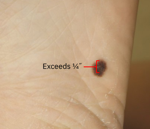 Illustration reference: OpenStax Clinical Nursing Skills Ch.21.2.
- Assess understanding of when to seek dermatology or primary-care evaluation.
- Reinforce that biopsy is required for definitive diagnosis when suspicious lesions are present.
Nursing Interventions
- Teach sunscreen and sun-protection behaviors as primary prevention.
- Teach sunscreen use with at least SPF 30 plus reapplication and broad-coverage technique during outdoor exposure.
- Teach exposure-timing controls: seek shade and limit direct sun during peak UV window (about 10 a.m. to 4 p.m.).
- Reinforce protective-clothing use (for example hats, sunglasses, and long sleeves/pants) during prolonged UV exposure.
- Teach patients to perform periodic mole surveillance using ABCDE criteria.
- Reinforce prompt evaluation when suspicious lesion change is identified.
- Prepare patients for likely lesion-directed procedures after diagnosis confirmation (for example curettage/cauterization, cryosurgery, excision, or Mohs surgery).
- Encourage annual dermatology skin checks for clients with prior skin-cancer history or frequently changing moles.
- In melanoma pathways, reinforce urgency because aggressive spread can require multimodal treatment in addition to excision.
- Teach that tanning beds are not a safe alternative to sun exposure and increase melanoma risk.
- Use clear shared decision-making language when discussing benefit-risk uncertainty of routine screening tests.
Delayed Evaluation Risk
Ignoring evolving mole changes can delay melanoma diagnosis and worsen prognosis.
Pharmacology
Prevention remains central, but treatment-stage choices become relevant after pathology confirmation. Provider plans may include local procedure-based removal, radiation in selected disease, and immunotherapy in advanced melanoma or selected advanced squamous-cell pathways.
Clinical Judgment Application
Clinical Scenario
A patient reports a mole that became darker, developed irregular borders, and increased in size over two months.
- Recognize Cues: Multiple ABCDE warning signs are present.
- Analyze Cues: Findings are concerning for possible melanoma progression.
- Prioritize Hypotheses: Urgent dermatologic evaluation is the priority.
- Generate Solutions: Provide immediate referral pathway and reinforce sun-protection measures.
- Take Action: Escalate for prompt specialist assessment and document lesion-change details.
- Evaluate Outcomes: Diagnostic delay is reduced and definitive management begins earlier if malignancy is confirmed.
Related Concepts
- integumentary-system - Structural and functional context for lesion pattern changes.
- benign-skin-tumors-and-lesions - Differential features that can mimic malignant lesions.
- cutaneous-lesion-procedures-and-postprocedure-care - Postbiopsy and postexcision care pathways including reconstruction and wound coverage.
- adult-preventive-screening-and-health-promotion - Secondary-prevention counseling framework across adult risk groups.
- well-care-anticipatory-guidance-and-immunization-across-the-lifespan - Preventive teaching workflow that can include skin-risk education.
Self-Check
- Which ABCDE finding should prompt the fastest referral urgency?
- Why is melanoma emphasized more strongly than basal cell carcinoma in warning-sign education?
- How do nurses combine primary and secondary prevention for skin-cancer risk reduction?