Eye Assessment Visual Acuity and Common Abnormalities
Key Points
- Eye assessment combines structural inspection, pupillary response checks, and visual acuity measurement.
- Snellen and Jaeger tools support standardized distance and near-vision baseline documentation.
- New pupillary asymmetry, sudden vision loss, papilledema-pattern cues, or painful red eye require rapid escalation.
- Reliable screening integrates external/internal anatomy, visual-field function, and reflex pathways rather than isolated single findings.
Pathophysiology
Vision depends on coordinated function of external eye structures, lens and retina, optic nerve pathways, and reflex control systems. Disruption at any level can alter acuity, fields, light response, or ocular comfort.
Progressive internal pathology may present with subtle early findings, so serial, objective assessments are essential for safe detection and referral. External-protection structures (eyelids/eyelashes/tear system/conjunctiva) and optical elements (cornea, lens, retina) work together with neural pathways (optic nerve, chiasm, tract, thalamic relay, visual cortex) to generate visual perception. Retinal photoreceptors support different functions: rods support low-light and peripheral vision, while cones support color discrimination and high-acuity central vision.
Pediatric visual development affects interpretation: full adult-level visual acuity is not typically reached until around age 7, and children younger than about 9 years have higher light/UV sensitivity than adults.
Classification
- Assessment domains: External-eye inspection, pupil size/reactivity, visual acuity (distance and near), and symptom-focused interview.
- Anatomy domains: External structures (brow, lids/lashes, conjunctiva, cornea, sclera, iris, pupil, lacrimal system) and internal structures (lens, ciliary muscle, vitreous, retina, optic nerve/disc).
- Visual-function domains: Central-detail vision versus peripheral-threat/motion detection.
- Reflex domains: Light, accommodation-convergence, blink-protective, and gaze-stabilizing reflexes.
- Common older-adult patterns: Presbyopia, cataracts, glaucoma, and macular degeneration.
- Macular degeneration subtype pattern: Dry (nonexudative) progression with drusen/scarring versus wet (exudative) progression with neovascular leakage, hemorrhage, and faster central-vision decline.
- Chronic-disease complication pattern: Diabetic retinopathy with progressive retinal vascular damage and preventable blindness risk.
- Abnormality groups: External inflammatory/structural findings and internal retinal/optic-nerve/lens disorders.
- Common external-abnormality examples: conjunctivitis, nasolacrimal-duct-obstruction, hordeolum, chalazion, ptosis, ectropion, and exophthalmos patterns.
- Common internal-abnormality examples: Pterygium, corneal abrasion/scar, cataract, retinal and optic-nerve disorder patterns.
- Environmental-irritant pattern: Chronic wind, ultraviolet-light, and dust exposure can contribute to pterygium progression.
- Pupil/optic-disc warning patterns: Persistent anisocoria, abnormal mydriasis or miosis, and papilledema-linked intracranial-pressure concern.
- Visual-abnormality spectrum: Refractive disorders (myopia, hyperopia, astigmatism, presbyopia), alignment/development disorders (strabismus, amblyopia), and symptom syndromes (diplopia, flashes/floaters, photophobia, night-vision decline, halos).
- Color-vision disorder pattern: Congenital or acquired color-deficiency syndromes with impaired shade discrimination.
- Amblyopia-risk pattern: Untreated refractive error asymmetry, visual-deprivation states, or strabismus can drive cortical suppression of the weaker eye and cause irreversible vision loss without early treatment.
- Blepharitis-pattern split: Acute ulcerative (often infectious) versus chronic meibomian/seborrheic inflammatory disease with recurrent flare profile.
- Chalazion pattern: Typically unilateral, painless, nonerythematous meibomian/Zeis gland blockage rather than primary acute infection.
- Urgency tiers: Chronic progressive change versus acute, vision-threatening or neurologic-associated change.
Nursing Assessment
NCLEX Focus
Do not treat visual acuity values in isolation; always integrate pain, pupil findings, and neurologic cues.
-
Assess visual symptoms: blurring, field loss, photophobia, pain, redness, discharge, and trauma history.
-
Collect focused exposure history including occupation, hobbies, and environmental irritants that increase ocular-surface injury risk.
-
Screen for symptom clusters that suggest structural progression: new flashes/floaters, central distortion or dark spots, night-vision decline, halos, or new double vision.
-
Inspect eyelids, conjunctiva, sclera, corneal clarity, and symmetry of ocular movement.
-
Use normal scleral baseline (typically white) to identify abnormal redness, swelling, or streaking.
-
Assess extraocular movement in all cardinal directions and correlate abnormalities with cranial-nerve III, IV, and VI function.
-
Test six cardinal fields of gaze with head stabilized and identify abnormal smooth pursuit, jerky tracking, or spontaneous/evoked nystagmus.
-
Assess pupils for equal size, shape, and light response; note anisocoria or abnormal dilation/constriction trends.
-
Use expected adult pupil-size context during interpretation (approximately 2-4 mm in bright light and 4-8 mm in darkness).
-
Check direct and consensual light response, then accommodation (constrict near, dilate distance) to identify pathway dysfunction.
-
Interpret abnormal pupil trends in context: unilateral/unequal size pattern, unexpected dilation, or unexpected constriction may reflect ocular, medication-related, or neurologic pathology.
-
Test convergence by moving a near target toward the bridge of the nose (about 2 inches) and confirming symmetric inward movement.
-
Measure visual acuity using standardized distance and near tools with consistent documentation method.
-
For distance acuity, use Snellen testing at 20 feet (6 meters) with usual corrective lenses and record fraction interpretation consistently (for example 20/40).
-
If corrective lenses are used during acuity screening, document results explicitly as corrected vision.
-
Use developmentally appropriate alternatives for non-reading children when Snellen letters are not valid (for example symbol/picture-based acuity methods).
-
For near acuity, use a Jaeger-style card at about 14-16 inches and document smallest comfortably readable line (for example J5 notation).
-
Include tonometry and funduscopic findings in the structured exam record when completed by the eye-care team, and escalate elevated IOP or optic-disc/macular abnormalities promptly.
-
Screen peripheral fields with confrontation testing across superior/inferior/temporal/nasal quadrants while central gaze is maintained.
-
Screen for common external-eye symptom patterns (for example diffuse red-itchy eye with discharge/crusting versus localized tender lid bump versus nonpainful gland blockage).
-
During routine inspection, treat yellow sclera as possible hepatobiliary dysfunction cue and sunken-eye appearance as possible dehydration cue.
-
In amblyopia concern, assess for squinting, persistent eye closure preference, head tilt, and side-favoring behavior.
-
In blepharitis concern, assess bilateral morning-predominant lid crusting, burning/foreign-body sensation, and meibomian secretion changes.
-
In recurrent chalazion concern, evert eyelids when indicated and screen for history of repeated lesions.
-
In suspected myopia, assess distance-vision complaints, squinting, frequent headaches, and behavior such as holding objects close.
-
In suspected hyperopia, assess near-vision blur/fatigue and contextual risks (for example age-related change, trauma, diabetes, and family history).
-
In strabismus concern, assess constant versus intermittent misalignment, diplopia/head-tilt compensations, and amblyopia-risk progression.
-
In color-vision concern, assess functional effect on shade discrimination and learning tasks and validate with color-plate testing patterns when available.
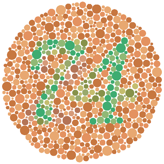 Illustration reference: OpenRN Nursing Health Promotion Ch.16.22.
 -
In periorbital infection concern, distinguish superficial periorbital swelling from orbital warning cues (painful or limited eye movement, reduced vision, fever, and deeper tissue involvement).
-
Screen for internal-structure risk patterns such as gradual peripheral-vision loss (glaucoma concern) and optic-disc edema findings (papilledema concern).
-
Correlate central-vision decline patterns with lens/macular pathology and peripheral-field loss patterns with glaucoma-risk progression.
-
In cataract concern, assess functional impact on reading, driving, and fall risk in addition to visual-acuity trends.
-
Assess risk context for major visual disorders (for example age, smoking, hypertension, and diabetes) and ensure follow-up eye-care referral continuity.
-
Review current medications for ocular risk contribution (for example corticosteroid exposure increasing glaucoma/cataract risk).
-
For clients with diabetes, verify annual retinal/eye examination status and reinforce early treatment pathways to prevent permanent vision loss.
Nursing Interventions
- Escalate sudden vision change, painful red eye, severe photophobia, or abnormal neurologic-associated pupillary findings.
- Escalate papilledema-pattern findings or acute pupil asymmetry with headache/neurologic change for urgent ICP-focused evaluation.
- Escalate new flashes/floaters, sudden diplopia, or rapidly worsening central/peripheral vision because retinal/optic-nerve pathology may be time-sensitive.
- Protect affected eye from additional trauma and avoid delay in specialist evaluation when red flags are present.
- Standardize exam setup (lighting, gaze target, one-eye-at-a-time method, corrective-lens use) to improve repeatability across reassessments.
- Validate discrepant findings by repeating focused exam and obtaining second-clinician verification when needed.
- Document appearance and function systematically: periorbital tissue, conjunctiva/cornea/iris, pupil size/reactivity, ocular movement quality, and near/far acuity results.
- Reinforce eye-protection, chronic-condition follow-up, and medication adherence for known ocular disease.
- Teach and reinforce routine annual eye examinations across adulthood, with earlier follow-up if new symptoms occur.
- Reinforce referral completion when outpatient or school screening identifies reduced acuity, color-vision deficits, or other abnormal vision results.
- Reinforce amblyopia-treatment adherence (corrective lenses, patching schedule, and prescribed atropine to unaffected eye) because delayed adherence can reduce visual-recovery potential.
- In blepharitis pathways, teach ongoing lid-hygiene routine (warm compresses, gentle lid cleansing, and cosmetic-trigger minimization) and escalate refractory disease.
- In chalazion pathways, reinforce warm-compress/eyelid-massage trial and avoid routine self-directed antibiotic use for noninfectious lesions.
- In hordeolum pathways, reinforce warm-compress routine (about 15 minutes, multiple times daily), gentle lid hygiene, and escalation for very large or nonresolving lesions.
- Escalate suspected orbital-extension cellulitis urgently and support imaging/referral workflows when periorbital infection severity is unclear.
Vision-Threatening Delay
Waiting on repeat routine checks when acute vision red flags are present can lead to irreversible visual loss.
Pharmacology
| Drug Class | Examples | Key Nursing Considerations |
|---|---|---|
| antiglaucoma-medications | Topical pressure-lowering drops | Confirm administration technique and monitor adherence and symptom change. |
| ophthalmic-antibiotics | Topical ocular antibiotics | Use for indicated infectious findings and monitor for worsening pain/redness. |
Clinical Judgment Application
Clinical Scenario
A patient reports sudden unilateral blurred vision with headache and unequal pupils on exam.
- Recognize Cues: Acute visual change plus abnormal pupillary pattern.
- Analyze Cues: Findings may represent urgent ocular or intracranial process.
- Prioritize Hypotheses: Immediate priority is prevention of permanent vision or neurologic injury.
- Generate Solutions: Trigger urgent escalation and focused reassessment.
- Take Action: Communicate red flags immediately and document objective exam findings.
- Evaluate Outcomes: Rapid referral supports timely diagnosis and treatment.
Related Concepts
- assisting-with-sensory-deficits - Supports day-to-day adaptation for persistent visual impairment.
- sensory-perception-and-reticular-activating-system - Connects visual input with perception and safety.
- documenting-and-reporting-data - Standardized charting improves trend interpretation.
- fall-prevention - Visual decline increases mobility and environmental hazard risk.
Self-Check
- Which eye findings require immediate escalation rather than routine follow-up?
- Why should pupillary findings be interpreted with neurologic context?
- How do standardized visual acuity methods improve patient safety over time?